Can Varicose Veins Cause a Blood Clot? DVT vs Superficial Clot
Category: Blogs
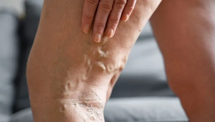
You felt something new — a painful, hard, red area along a varicose vein in your leg. Or maybe your whole calf swelled up and started aching in a way that felt different and more serious than before.
Yes, varicose veins can cause blood clots — and knowing the difference between a superficial clot and a deep vein clot could be the most important thing you read today.
If you're concerned about varicose veins blood clot treatment in Pune, Lokmanya Hospitals provides specialist vascular care with advanced duplex imaging and expert surgical management for all stages of venous disease.
Key Takeaways — In This Article You Will Learn:
- Why varicose veins increase your risk of blood clot formation
- The clear difference between a superficial clot and a dangerous DVT
- Which symptoms require emergency care — and which can wait
- How clots are diagnosed and treated in 2025
- What you can do to prevent clots if you have varicose veins
Can Varicose Veins Actually Cause a Blood Clot?
Yes — varicose veins directly increase the risk of blood clot formation, and this is one of the most underappreciated complications of untreated venous disease.
Why Varicose Veins Create the Right Conditions for Clotting?
In medicine, there is a concept called Virchow's Triad — three conditions that together create the ideal environment for a blood clot to form.
These three conditions are: slow or stagnant blood flow, damage to the vein wall, and blood that clots more easily than normal.
Varicose veins tick all three boxes.
Blood pools in the stretched, damaged vein instead of flowing smoothly upward. The vein wall itself is structurally abnormal from years of pressure. And in many patients, additional factors like dehydration, prolonged sitting, or illness tip the balance toward clot formation.
If you are still learning about the condition itself, our earlier guide on varicose veins — their symptoms, stages, and early warning signs — provides useful background.
What Superficial Thrombophlebitis Actually Is?
When a blood clot forms inside a varicose vein — a vein that sits just under the skin — the condition is called superficial thrombophlebitis.
"Thrombo" means clot. "Phlebitis" means inflammation of the vein.
The clot and the inflammation around it are what cause the redness, warmth, hardness, and pain you feel along the vein.
This is different from a deep vein clot — but it is not something to dismiss.
How Common Is This Complication?
Research estimates that approximately 20–59% of patients with varicose veins will develop superficial thrombophlebitis at some point if their venous disease is left untreated.
It is far more common than most patients — and some doctors — communicate clearly. This is a real complication of a real progressive disease.
What Is the Difference Between a Superficial Clot and DVT?
This is the question that matters most — because the answer determines how urgently you need care.
Where Each Type of Clot Forms
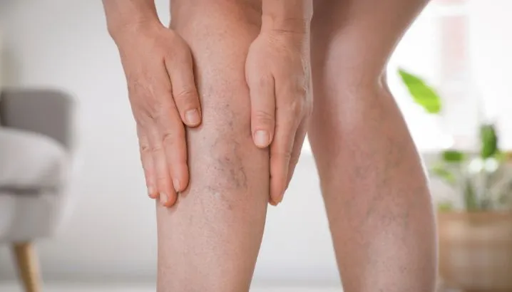
Superficial thrombophlebitis forms in veins that sit just below the skin — the same veins you can see and feel when varicose veins are present.
Deep vein thrombosis (DVT) forms in the deep veins that run inside the muscle, hidden from the surface. These veins carry far larger volumes of blood and are directly connected to the lungs via the bloodstream.
Why the Location Changes Everything?
A clot in a superficial vein is painful and needs treatment — but it does not directly threaten your lungs or life in the way a DVT can.
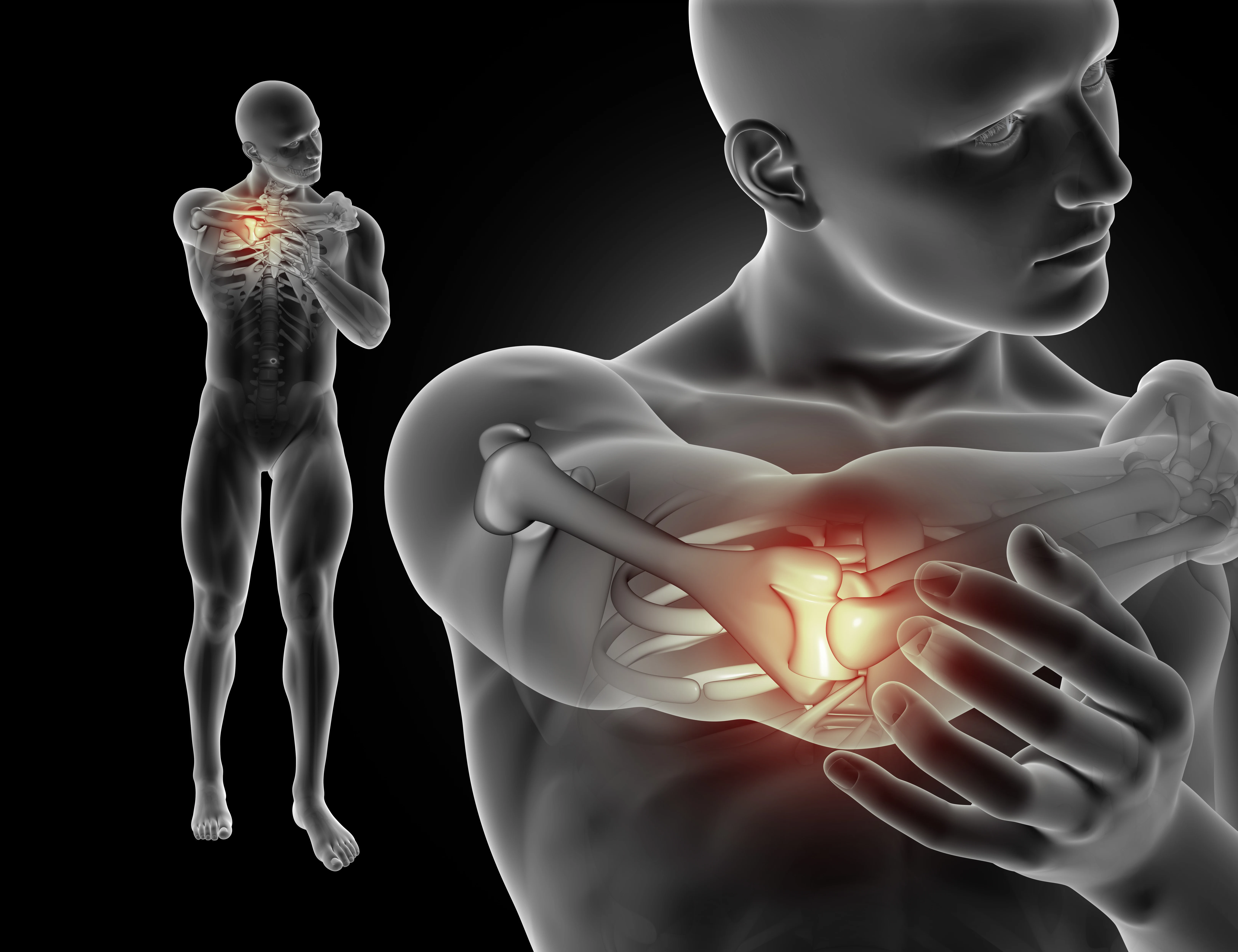
A DVT carries the risk of pulmonary embolism — where part of the clot breaks off, travels through the bloodstream, and blocks a blood vessel in the lung. This is a life-threatening emergency.
This is why distinguishing between the two is not a matter of preference — it is a matter of safety.
Superficial Thrombophlebitis vs DVT — At a Glance
Because this topic is a direct comparison between two distinct conditions, a structured overview genuinely helps patients understand the difference:
Feature | Superficial Thrombophlebitis | Deep Vein Thrombosis (DVT) |
| Where it forms | Just under the skin, in varicose veins | Deep inside the leg muscles |
| Visible on surface? | Yes — red, hard, tender vein visible | Usually not visible |
| Main symptoms | Localised pain, redness, warmth, hardness along vein | Whole-leg swelling, diffuse aching, heaviness |
| Skin colour change | Redness over the vein | Possible bluish or pale discolouration of whole leg |
| Risk of lung clot (PE)? | Low — unless it extends to deep system | High — this is the primary danger |
| Diagnosis | Duplex ultrasound | Duplex ultrasound + D-dimer blood test |
| Treatment | Anti-inflammatories, compression, anticoagulation if near deep veins | Anticoagulation (blood thinners), sometimes hospitalisation |
| Urgency | Seek care within 24–48 hours | Seek emergency care same day |
What Does a Blood Clot in a Varicose Vein Feel Like?
Symptoms of Superficial Thrombophlebitis
The presentation is usually localised and relatively specific.
You will typically feel a firm, cord-like hardness along the path of a varicose vein. The skin over it turns red and feels warm to touch. The area is tender when pressed.
Unlike general varicose vein discomfort, this pain is more focused, more intense, and does not improve with elevation the way ordinary vein aching does.
Symptoms That Suggest DVT Instead
DVT symptoms are different in character and distribution.
Sudden swelling of the entire calf or leg — not just around one vein — is the most important warning sign. The leg may feel heavy, tight, and ache deeply rather than along a specific surface line.
Some people develop a bluish or pale discolouration of the leg. A few develop no symptoms at all — which is one reason DVT is genuinely dangerous.
If your whole leg swells suddenly, seek emergency evaluation the same day. Do not wait.
The Overlap That Makes Self-Diagnosis Risky
Here is the honest clinical truth: superficial and deep clots can coexist in the same leg at the same time, and symptoms can overlap.
A patient who has obvious superficial thrombophlebitis along a visible vein may also have a silent DVT in the deeper system. This is why imaging — not symptom assessment alone — is essential for any new leg clot.
When Can a Superficial Clot Become a Deep Vein Clot?
The Extension Pathway
Clots do not always stay where they form. Under the right conditions, a superficial clot can grow — extending along the vein toward the point where the superficial and deep venous systems connect.
Once a clot crosses from the superficial into the deep system, the clinical situation changes entirely. It is now a DVT with all the associated risks.
Which Patients Face the Highest Extension Risk?
Extension risk is higher in patients with large clot burden, rapidly enlarging clots, clots in the thigh rather than the calf, or patients with additional clotting risk factors such as recent surgery, cancer, pregnancy, or a known clotting disorder.
Patients on oral contraceptives or hormone therapy also face elevated risk. These groups need more urgent and more aggressive management.
The Saphenofemoral Junction — The Critical Anatomy
This is the anatomical detail almost no patient-facing content explains — and it is the most important thing to understand about superficial clot extension.
The saphenofemoral junction is the point where the great saphenous vein (the main superficial leg vein) joins the deep femoral vein in the groin.
When a clot in the saphenous vein extends to within 3cm of this junction, it is considered at immediate risk of entering the deep system. This is a vascular emergency requiring anticoagulation regardless of whether the patient has DVT symptoms.
Duplex ultrasound is the only way to know how close your clot is to this junction. This is why clinical examination alone is never sufficient.
How Is a Leg Clot Diagnosed?
Why Clinical Examination Is Not Enough?
A doctor can identify signs highly suggestive of superficial thrombophlebitis by examination alone.
But examination cannot tell you whether a DVT is also present, how large the clot is, or how close it is to the deep venous system. These are the questions that drive treatment decisions — and they require imaging.
Duplex Ultrasound — The Definitive Tool
Duplex ultrasound is the gold standard investigation for any suspected leg clot. It combines real-time imaging of the veins with blood flow assessment.
It maps exactly where the clot begins and ends, whether it has reached the saphenofemoral junction, and whether deep veins are involved. It is painless, non-invasive, and results are available immediately.
Any patient with a new, painful, firm area along a leg vein should have a duplex scan — not just a clinical review.
D-Dimer — Useful But Not Definitive
The D-dimer blood test measures a protein fragment released when a blood clot breaks down. An elevated result suggests a clot may be present somewhere in the body.
However — and this is the limitation most patients are never told — D-dimer has a very high false-positive rate. Pregnancy, infection, recent surgery, and even simple inflammation can elevate it.
A positive D-dimer does not diagnose DVT. It indicates that further imaging is needed. A normal D-dimer in a low-risk patient can help rule out DVT — but it is not the final word on its own.
How Are Superficial Clots and DVT Treated?
Managing Superficial Thrombophlebitis
For small, localised clots well away from the deep venous system, treatment focuses on reducing inflammation and preventing extension.
NSAIDs (anti-inflammatory medications like ibuprofen or diclofenac) reduce pain and local inflammation. Compression stockings support venous return and reduce further pooling. Keeping active — walking regularly — is encouraged to maintain calf muscle pump function.
For clots within 3cm of the saphenofemoral junction or extending rapidly, anticoagulation (blood-thinning medication) is required — typically a low molecular weight heparin injection or a direct oral anticoagulant for 45 days, as supported by the CALISTO trial evidence.
Anticoagulation for DVT
DVT requires anticoagulation — blood-thinning medication — to prevent clot extension and pulmonary embolism.
Modern oral anticoagulants (DOACs) such as rivaroxaban or apixaban have largely replaced older warfarin-based regimens for most patients. They are taken as tablets, require no regular blood test monitoring, and are highly effective.
Treatment duration is typically 3–6 months for a first DVT provoked by a clear cause, and longer for unprovoked clots or recurrent events.
When Varicose Vein Treatment Prevents Recurrence?
Treating the underlying varicose veins — through EVLT or sclerotherapy — removes the structural environment in which clots form. Patients who have had superficial thrombophlebitis and then have their varicose veins treated have a significantly lower recurrence risk than those whose veins are left untreated.
If you are exploring your options, Lokmanya Hospitals offers a full range of varicose vein treatments in Pune — from Endovenous Laser Ablation (EVLA) and Radiofrequency Ablation to VenaSeal and sclerotherapy — all delivered as walk-in, walk-out procedures.
Clot treatment manages the acute problem. Vein treatment addresses the cause.
The Massage Myth — Please Read This Carefully
Do not massage a leg where you suspect a blood clot.
This is one of the most common and potentially dangerous things patients do — and no competitor content directly addresses it.
Massaging a clotted vein can dislodge the clot or cause it to fragment and travel through the bloodstream. If the clot reaches the lungs, the result is a pulmonary embolism.
If your leg is painful, firm, swollen, or red — rest, elevate gently, and seek medical evaluation. Do not apply pressure or massage the area.
If you have varicose veins in Pimpri Chinchwad and have noticed a new hard, red, painful area along a vein — or sudden leg swelling — the vascular team at Lokmanya Hospitals, Chinchwad is equipped to assess and treat you with same-day duplex imaging. Visit lokmanyahospitals.com to book an urgent vascular consultation.
Can You Prevent Blood Clots If You Have Varicose Veins?
Compression and Movement as Daily Protection
Medical-grade compression stockings reduce venous pooling and lower the risk of clot formation in patients with known varicose veins.
Regular walking — even short walks every 30–45 minutes during a long workday — activates the calf muscle pump and keeps blood moving.
These are not cures. They are daily risk-reduction measures that work alongside, not instead of, proper medical treatment.
High-Risk Periods That Need Extra Caution
Certain situations dramatically increase clotting risk in patients who already have varicose veins.
Long-haul flights or car journeys over 4 hours, post-surgical immobility, hospital admissions, and pregnancy all compound existing venous disease risk.
During these periods: wear compression stockings, stay hydrated, move your ankles regularly if seated, and discuss thromboprophylaxis with your doctor if surgery is planned.
When Treating the Veins Reduces Long-Term Clot Risk?
Definitive varicose vein treatment — EVLT, sclerotherapy, or surgical intervention — does not just improve symptoms. It removes the abnormal venous architecture that creates the conditions for clotting.
Multiple studies confirm that patients who undergo successful varicose vein treatment have lower rates of superficial thrombophlebitis recurrence. Treating the veins is the most durable form of clot prevention available to this patient group.
Final Thoughts
Varicose veins are not a cosmetic problem with cosmetic risks. They can cause blood clots — and those clots can range from painful but manageable to life-threatening, depending on where they form and how quickly they are identified.
The difference between a superficial clot and a DVT is not always obvious from symptoms alone. Duplex ultrasound is the only reliable way to tell them apart, assess clot extent, and guide treatment safely.
The key messages: don't ignore new pain or hardness along a varicose vein; never massage a suspected clot; seek same-day emergency care for sudden whole-leg swelling; and consider treating your varicose veins definitively to reduce your long-term risk.
Help is available — and the earlier you act, the simpler the treatment required.
Concerned about a painful or swollen leg? Don't wait for symptoms to worsen. The vascular specialists at Lokmanya Hospitals in Pune offer expert assessment for DVT symptoms in legs and superficial thrombophlebitis, with same-day duplex imaging and personalised treatment planning. Visit lokmanyahospitals.com to book your consultation today.
Frequently Asked Questions
Can a superficial blood clot spread to a deep vein?
Yes — and this is exactly why superficial clots need proper evaluation, not just rest and painkillers. A clot that extends to within 3cm of the saphenofemoral junction — the point where the superficial and deep venous systems connect — is at immediate risk of becoming a DVT. Only a duplex ultrasound can tell you how close your clot is to that junction.
Is it safe to walk with a superficial blood clot in my leg?
Gentle walking is generally safe and encouraged for superficial thrombophlebitis — it activates the calf muscle pump and helps maintain blood flow. However, if a DVT has not been ruled out by ultrasound, strenuous activity should wait until imaging is complete. Never massage the affected area regardless of which type of clot is suspected.
What happens if DVT is not treated?
Untreated DVT carries a significant risk of pulmonary embolism — where part of the clot breaks off and travels to the lungs, potentially causing sudden breathlessness, chest pain, and in severe cases, death. Long-term, untreated DVT also causes permanent damage to the deep vein valves, leading to a condition called post-thrombotic syndrome — chronic leg swelling, pain, and skin changes that can last for years.
How long does it take for a superficial blood clot to go away?
With appropriate treatment — anti-inflammatories, compression, and anticoagulation where indicated — most superficial clots resolve over 2–6 weeks. The hardness along the vein often takes longer to fully soften than the pain and redness take to settle. Follow-up duplex imaging confirms resolution and checks that the clot has not extended during the recovery period.
Can I fly if I have varicose veins and a history of blood clots?
Flying is not absolutely prohibited, but it requires preparation. Long flights combine immobility, dehydration, and reduced cabin pressure — all of which increase clotting risk in patients with varicose veins or a prior clot history. Wear properly fitted compression stockings for the flight, stay well hydrated, and perform regular ankle exercises. Discuss anticoagulation prophylaxis with your doctor before any flight over 4 hours if you have had a prior DVT.