X-Ray vs MRI: Understanding Spine Investigations
Category: Orthopedics
Each spinal segment consists of two key components: the vertebral bones and the nerves. Although they function together, they are evaluated differently during medical investigations because each imaging method highlights specific structures.
An X-ray primarily focuses on the bony structures of the spine. It helps doctors assess whether the vertebrae are properly aligned, whether there is abnormal movement, a reduction in vertebral height, or the presence of fractures. These structural changes are clearly visible on X-rays and make them an important first-line investigation in spine-related complaints.
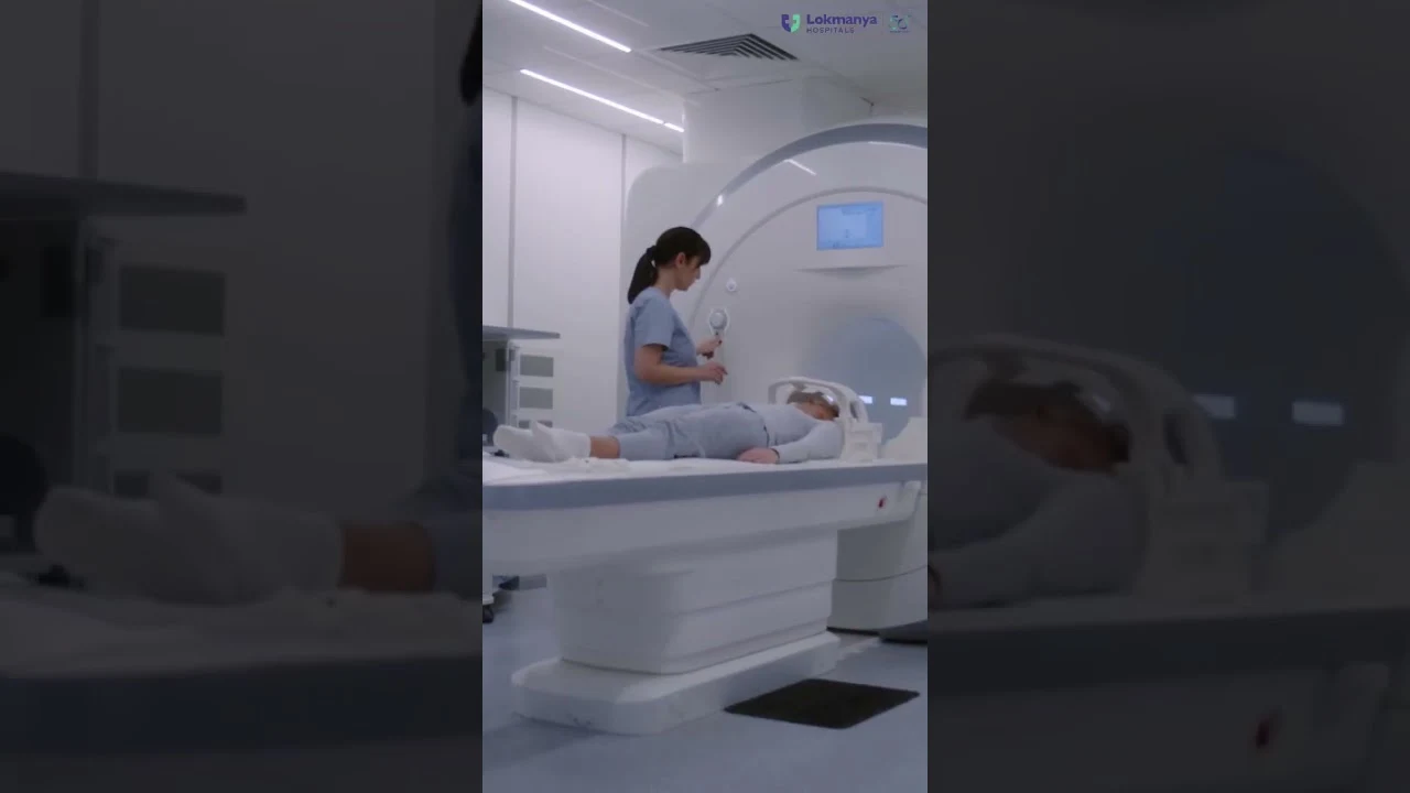
However, when the concern involves nerves or soft tissues, an X-ray alone is not sufficient. Conditions such as nerve compression, a slipped or torn disc, pressure caused by disc herniation, tumors, or other soft tissue pathologies cannot be adequately visualized on an X-ray. In such cases, an MRI (Magnetic Resonance Imaging) scan becomes essential.
In most cases, the diagnostic approach begins with an X-ray to rule out fractures or spinal instability. If symptoms or findings suggest possible nerve involvement, an MRI is then advised for a more detailed evaluation. In some situations, patients may already have an MRI done first, and an X-ray is later recommended to assess spinal movement and alignment. This combined approach helps provide a complete picture of the spine’s condition.
Why both tests matter
Both X-rays and MRI scans play a crucial role in diagnosing spinal disorders. While X-rays assess bone structure and movement, MRI scans provide detailed insight into nerves, discs, and soft tissues. Together, they ensure accurate diagnosis and appropriate treatment planning for spine-related conditions.